
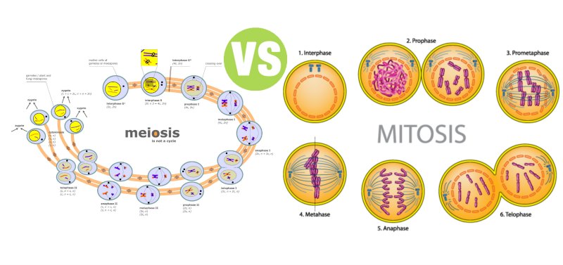
What is the Difference Between Mitosis and Meiosis?
Summary: Difference Between Mitosis and Meiosis is that mitosis is divided into four phases: prophase, metaphase, anaphase, and telophase. In a dividing cell, however, the process is actually continuous, with each phase smoothly flowing into the next. While a type of cell division called meiosis (Gr. meiosis, dimunition) occurs in specialized cells of the ovaries and testes and reduces the number of chromosomes to the haploid (1N) number.
Mitotic Cell Division
Cell division occurs in all animals during growth and repair processes. Walther Flemming while studying the dividing cells of orchid plant used the term “Mitosis” meaning division of parent nucleus into daughter nuclei. Cells divide in two basic stages: Mitosis is division of the nucleus, and cytokinesis (Gr. kytos, hollow vessel + kinesis, motion) is division of the cytoplasm. Between divisions (interphase), the cell must grow and carry out its various metabolic processes. The Cell Cycle is that period from the time a cell is produced until it completes Mitosis. Karyokinesis (division of nucleus) and cytokinesis (division of cytoplasm) make up the M (mitotic) phase of the cell cycle.
G1
The G1 (First growth of gap) phase represents the early growth phase of the cell. During the S (DNA Synthesis) phase, growth continues, but this phase also involves DNA Replication. The G2 (Second growth of gap) phase prepares the cell for the division. It includes replication of the Mitochondria and other organelles, synthesis of microtubules and protein that will make up the mitotic spindle fibers, and chromosome condensation.
The M (Mitotic) phase includes events associated with partitioning chromosomes between two daughter cells and the division of the cytoplasm (cytokinesis).Mitotic cell division, is a type of indirect cell division by which the parent cell divides into daughter cells, in such a way that the number of chromosomes remain identical as in parent cell.
Interphase (Replicating the Hereditary Material)
Interphase (L. inter, between) (includes the G1, S, and G2 phases) typically occupies about 90% of the total cell cycle. It is the period during which the normal activities of the cell take place. Interphase also sets the stage for cell division because DNA replication is completed during the S phase of interphase. Before a cell divides, an exact copy of the DNA is made. This process is called replication, because the double-stranded DNA makes a replica or duplicate, of itself. Replication is essential to ensure that each daughter cell receives the same genetic material as is present in the parent cell. The result is a pair of sister chromatids. A chromatid is a copy of a chromosome produced by replication. Each chromatid attaches to its other copy, or sister, at a point of constriction called a centromere.
The centromere is a specific DNA sequence of about 220 nucleotides and has a specific location on any given chromosome. Bound to each centromere is a disk of protein called a kinetochore, which eventually is an attachment site for the microtubules of the mitotic spindle. There is a concept that during interphase, the enzymes needed for the removal of kinetochore are absent, as a result the centromeric part of chromosome is unable to replicate during interphase. So, duplication of centromere takes place during anaphase.
G2
As the cell cycle moves into the G2 phase the chromosomes begin condensation. During the G2 phase, the cell also begins to assemble the structures that it will later use to move the chromosomes to opposite poles (ends) of the cell. For example, centrioles replicate, and there is extensive synthesis of the proteins that make up the microtubules. The enzymes synthesized during S-phase catalyze this G2-phase.
Cell Cycle Control
It is evident that nuclear events do not control cell cycle. Instead a set of chemical reactions in cytoplasm controls cell division. The mechanism of cell cycle control involves cyclin protein activating and inactivating factors along with maturation-promoting factor (MPF).
During interphase cyclin accumulates until the rate of MPF activation (by cyclin) exceeds the rate of cyclin destruction by an inactivating enzyme. As a result active maturation-promoting factor (MPF) accumulates leading to a series of modifications.
MITOSIS
Mitosis is divided into four phases: prophase, metaphase, anaphase, and telophase. In a dividing cell, however, the process is actually continuous, with each phase smoothly flowing into the next.

Prophase
The first phase of mitosis, prophase (Gr. pro, before phase), begins when chromosomes become visible with the light microscope as threadlike structures. The nucleoli and nuclear envelope begin to break up, and the two centriole pairs move apart. By the end of prophase, the centriole pairs are at opposite poles of the cell. The centrioles radiate an array of microtubules called asters (L. aster, little star), which brace each centriole against the plasma membrane.
Between the centrioles, the microtubules form a spindle of fibers that extends from pole to pole. The asters, spindle, centrioles, and microtubules are collectively called the mitotic spindle (or mitotic apparatus). As prophase continues, a second group of microtubules grows out from the kinetochore to the poles of the cell. These kinetochore microtubules connect each sister chromatid to the poles of the spindle.
Metaphase
As the dividing cell moves into metaphase (Gr. Meta, after phase), the chromatids (replicated chromosomes) begin to align in the center of the cell, along the spindle equator. Toward the end of metaphase, the centromeres divide and detach the two sister chromatids from each other, although the chromatids remain aligned next to each other. After the centromeres divide, the sister chromatids are considered full-fledged chromosomes (called daughter chromosomes).
Anaphase
During anaphase (Gr. ana, back again phase), the shortening of the microtubules in the mitotic spindle pulls each daughter chromosome apart from its copy and toward its respective pole. Anaphase ends when all the daughter chromosomes have moved to the poles of the cell. Each pole now has a complete, identical set of chromosomes.
Telophase
Telophase (Gr. telos, end phase) begins once the daughter chromosomes arrive at the opposite poles of the cell. During telophase, the mitotic spindle disassembles. A nuclear envelope reforms around each set of chromosomes, which begin to uncoil for gene expression, and the nucleolus is resynthesized. The cell also begins to pinch in the middle. Mitosis is over, but cell division is not.
Cytokinesis: (partitioning the cytoplasm)
The final phase of cell division is cytokinesis, in which the cytoplasm divides. Cytokinesis usually starts sometime during late anaphase or early telophase. A contracting belt of microfilaments called the contractile ring pinches the plasma membrane to form the cleavage furrow. The furrow deepens, and two new, genetically identical daughter cells form.
Important Aspects Of Mitosis
- Maintains a constant number of chromosomes through successive cell divisions. That’s why each cell in our body has 46 chromosomes, like fertilized egg (with 46 chromosomes) which give rise to all cells of our body by repeated divisions.
- Mitosis divides bivalent chromosomes (homologous chromosome pair) into monovalent, thus allowing the replication of DNA and duplication of chromosomes during interphase essential for development.
- Mitosis allows the conservation of genotype (the specific allele combinations situated on corresponding chromosomes, determining a specific trait of an individual) of zygote.
- Mitosis helps to keep the nucleus and cytoplasm ratio constant, vital for homeostatic functioning (the ability of body to seek and maintain a condition of equilibrium within its internal environment, even facing external changes) of the cells.
- Different mitotic rates of different cell families help in differentiation and organogenesis (the formation and development 0f animals and plants organs)
More Confusing Biological Terms
Difference Between Dementia and Alzheimer’s
Difference Between Sex and Gender
Difference Between HMO and PPO
Difference Between DNA and RNA
Difference Between Sexual and Asexual Reproduction
Meiosis
Sexual reproduction requires a genetic contribution from two different sex cells. Egg and sperm cells are specialized sex cells called gametes (Gr. gamete, wife; gametes, husband). In animals, a male gamete (sperm) unites with a female gamete (egg) during fertilization to form a single cell called a zygote (Gr. zygotos, yoked together). The fusion of gametes is called syngamy (Gr. gamos, marriage). The zygote is the first cell of the new animal. Each of the two gametes (haploid mature female or male germ cells) contributes half of the genetic information to the zygote.
To maintain a constant number of chromosomes in the next generation, animals that reproduce sexually must produce gametes with half the chromosome number of their ordinary body cells (called somatic cells). All of the cells in the bodies of most animals, except for the egg and sperm cells, have the diploid (2N) number of chromosomes. A type of cell division called meiosis (Gr. meiosis, dimunition) occurs in specialized cells of the ovaries and testes and reduces the number of chromosomes to the haploid (1N) number. The nuclei of the two gametes combine during fertilization and restore the diploid number.
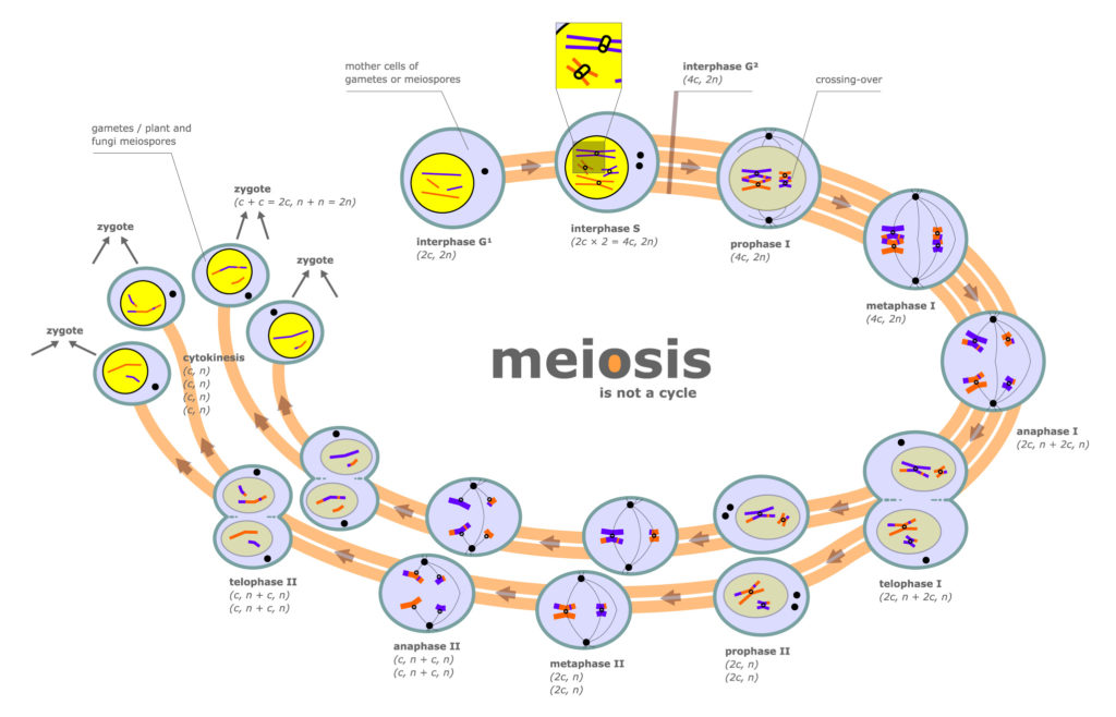
Meiosis begins after the G2 phase in the cell cycle—after DNA replication. Two successive nuclear divisions, designated meiosis I and meiosis II, take place. The two nuclear divisions of meiosis result in four daughter cells, each with half the number of chromosomes of the parent cell. Moreover, these daughter cells are not genetically identical. Like mitosis, meiosis is a continuous process, and biologists divide it into the phases that follow only for convenience.
The First Meiotic Division
In prophase I; chromatin (the chromosomes of all organisms other than bacteria contain a material containing protein DNA and RNA) folds and chromosomes become visible under a light microscope (figure 3.6a). Because a cell has a copy of each type of chromosome from each original parent cell, it contains the diploid number of chromosomes. Homologous chromosomes (homologues) carry genes for the same traits, are the same length, and have a similar staining pattern, making them identifiable as matching pairs.
During prophase I, homologous chromosomes line up side-by-side in a process called synapsis (Gr. synapsis, conjunction), forming a tetrad of chromatids (also called a bivalent). The tetrad thus contains the two homologous chromosomes, each with its copy, or sister chromatid. A network of protein and RNA is laid down between the sister chromatids of the two homologous chromosomes. This network holds the sister chromatids in a precise union so that each gene is directly across from its sister gene on the homologous chromosome.
Synapsis
Synapsis also initiates a series of events called crossing over, whereby the non-sister chromatids of the two homologous chromosomes in a tetrad exchange DNA segments (figure 3.7). This process effectively redistributes genetic information among the paired homologous chromosomes and produces new combinations of genes on the various chromatids in homologous pairs. Thus, each chromatid ends up with new combinations of instructions for a variety of traits. Crossing-over is a form of genetic recombination and is a major source of genetic variation in a population of a given species.
Metaphase
In metaphase I, the microtubules form a spindle apparatus just as in mitosis. However, unlike mitosis, where homologous chromosomes do not pair, each pair of homologues lines up in the center of the cell, with centromeres on each side of the spindle equator
Anaphase
Anaphase I begins when homologous chromosomes separate and begin to move toward each pole. Because the orientation of each pair of homologous chromosomes in the center of the cell is random, the specific chromosomes that each pole receives from each pair of homologues are also random
Meiotic Telophase
Meiotic telophase I is similar to mitotic telophase. The transition to the second nuclear division is called interkinesis. Cells proceeding through interkinesis do not replicate their DNA. After a varying time period, meiosis II occurs.
Second Meiotic Division
The second meiotic division (meiosis II) resembles an ordinary mitotic division, except the number of chromosomes has been reduced by half. The phases are prophase II, metaphase II, anaphase II, and telophase II. At the end of telophase II and cytokinesis, the final products of these two divisions of meiosis are four new “division products.” In most animals, each of these “division products” is haploid and may function directly as a gamete (sex cell).
EVENTS OF SECOND MITOTIC DIVISION ARE AS FOLLOWS:
Prophase-II:
A spindle apparatus is formed and the chromosomes move towards the metaphase-II plate.
Metaphase-II:
The chromosomes align on the metaphase plate in mitosis-like fashion, with the kinetochores of sister chromatids of each chromosome pointing toward opposite poles.
Anaphase-II:
The centromeres of sister chromatids finally separate and the sister chromatids of each pair are now considered as individual chromosomes. Now they move toward the opposite p0les of the cell.
Telophase-II and Cytokinesis:
Nuclei begin to form at opposite poles of the cell and cytokinesis occurs. There are now four daughter cells, each with the haploid number of chromosomes.
More Health Like Confusing Terms:
Difference Between Apoptosis and Necrosis
Difference Between Lysosomes and Peroxisomes
Difference Between Cell and Tissue
Difference Between Enamel and Cementum
Spermatogenesis and Oogenesis
The result of meiosis in most animals is the formation of sperm and egg cells. Spermatogenesis produces mature sperm cells and follows the sequence previously described. All four products of meiosis often acquire a flagellum for locomotion and a cap-like structure that aids in the penetration of the egg. Oogenesis produces a mature ovum or egg. Oogenesis differs from spermatogenesis in that only one of the four meiotic products develops into the functional gamete. The other products of meiosis are called polar bodies and eventually disintegrate. In some animals the mature egg is the product of the first meiotic division and only completes meiosis if it is fertilized.
What is the Difference Between Spermatogenesis and Oogenesis
They differ in three ways:
Meiotic Division
- During oogenesis, cytokinesis is unequal because almost all cytoplasm is monopolized by a single daughter cell known as secondary oocyte. This goes to form ovum and the other products of meiosis called polar bodies, degenerate. Whereas, in spermatogenesis all four products of meiosis-I and meiosis-II develop into mature sperms.
- The cells from which sperms develop continue to divide by mitosis throughout the male reproductive years. In females at the time of birth ovary already contains all the cells which later on, develop into eggs.
- Oogenesis, has long resting periods as compared to spermatogenesis in which mature sperms are produced from precursor cells in an uninterrupted sequence.
Similarities and Differences Between Mitosis and Meiosis
MITOSIS
An equational division which separates sister chromatids.
One division per cycle, i.e one cytoplasmic division per chromosomal division.
Chromosomes do not synapse; no chiasmata (point of contact between paired chromosomes) formation which means no genetic exchange occurs between homologous chromosomes.
Two products (daughter cells) are produced per cycle.
Genetic material of mitotic products is identical.
Chromosome number of daughter cell is identical to the chromosome number of mother cell.
Mitotic products are usually capable of undergoing additional mitotic divisions.
Mitotic division occurs in almost all somatic cells (all cells other than reproductive cells).
This division begins at the zygote stage and continues throughout the life of an organism.
MEIOSIS
First stage is a reduction division which separates homologous chromosomes at Anaphase-I; sister chromatids separate in an equational division at anaphase-II.
Two divisions per cycle, i.e two cytoplasmic divisions, one followed by reductional chromosomal division and the other followed by equational chromosomal division.
Chromosomes synapse and form chiasmata; genetic exchange occurs between homologous chromosomes.
Four cellular products (gametes) produced per cycle.
Genetic material of meiotic products is not identical; centromeres may be replicas of either maternal or paternal centromeres in varying combinations.
Chromosome number of meiotic products is half as compared to the chromosome number of mother cell.
Meiotic products cannot undergo another meiotic division (Although it may undergo mitotic division).
Meiotic division only occurs in specialized cells called germ cells (cells which develop into gametes).
Occurs only when higher organisms reach maturity.


Leave a Comment
You must be logged in to post a comment.