
Ribosomes vs Lysosomes
Summary: Difference Between Ribosomes and Lysosomes is that Ribosomes are the organelles without limiting membrane. These organelles are granular and small dot-like structures with a diameter of 15 nm. While Lysosomes are the membrane-bound vesicular organelles found throughout the cytoplasm.

RIBOSOMES
Ribosomes are the organelles without limiting membrane. These organelles are granular and small dot-like structures with a diameter of 15 nm. Ribosomes are made up of 35% of proteins and 65% of ribonucleic acid (RNA). RNA present in ribosomes is called ribosomal RNA (rRNA). Ribosomes are concerned with protein synthesis in the cell.
Types of Ribosomes
Ribosomes are of two types:
- Ribosomes that are attached to rough endoplasmic reticulum
- Free ribosomes that are distributed in the cytoplasm.
Functions of Ribosomes
Ribosomes are called ‘protein factories’ because of their role in the synthesis of proteins. Messenger RNA (mRNA) carries the genetic code for protein synthesis from nucleus to the ribosomes. The ribosomes, in turn arrange the amino acids into small units of proteins. Ribosomes attached to rough endoplasmic reticulum are involved in the synthesis of proteins such as the enzymatic proteins, hormonal proteins, lysosomal proteins and the proteins of the cell membrane.
Free ribosomes are responsible for the synthesis of proteins in hemoglobin, peroxisome and mitochondria.
Lysosomes
Lysosomes are the membrane-bound vesicular organelles found throughout the cytoplasm. The lysosomes are formed by Golgi apparatus. The enzymes synthesized in rough endoplasmic reticulum are processed and packed in the form of small vesicles in the Golgi apparatus. Then, these vesicles are pinched off from Golgi apparatus and become the lysosomes. Among the organelles of the cytoplasm, the lysosomes have the thickest covering membrane. The membrane is formed by a bilayered lipid material. It has many small granules which contain hydrolytic enzymes.
Types of Lysosomes
Lysosomes are of two types:
Primary lysosome, which is pinched off from Golgi apparatus. It is inactive in spite of having hydrolytic enzymes.
Secondary lysosome, which is the active lysosome. It is formed by the fusion of a primary lysosome with phagosome or endosome.
Functions of Lysosomes
Lysosomes are often called ‘garbage system’ of the cell because of their degradation activity. About 50 different hydrolytic enzymes, known as acid hydroxylases are present in the lysosomes, through which lysosomes execute their functions.
Important Lysosomal Enzymes
- Proteases, which hydrolyze the proteins into amino acids
- Lipases, which hydrolyze the lipids into fatty acids and glycerides
- Amylases, which hydrolyze the polysaccharides into glucose
- Nucleases, which hydrolyze the nucleic acids into mononucleotides.
Mechanism of Lysosomal Function
Lysosomal functions involve two mechanisms:
- Heterophagy: Digestion of extracellular materials engulfed by the cell via endocytosis
- Autophagy: Digestion of intracellular materials such as worn-out cytoplasmic organelles.
More Confusing Differences in Biology:
Difference Between Secretory Vesicles and Lysosomes
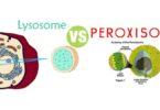
Difference Between Peroxisomes and Lysosomes
Difference Between Mitosis and Meiosis
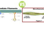
Difference Between Microtubules and Microfilaments
Difference Between Apoptosis and Necrosis
Difference Between Microtubules and Intermediate Filaments
Specific Functions of Lysosomes
1. Degradation of Macromolecules
Macromolecules are engulfed by the cell by means of endocytosis (phagocytosis, pinocytosis or receptormediated endocytosis. The macromolecules such as bacteria, engulfed by the cell via phagocytosis are called phagosomes or vacuoles. The other macromolecules taken inside via pinocytosis or receptor-mediated endocytosis are called endosomes. The primary lysosome fuses with the phagosome or endosome to form the secondary lysosome. The pH in the secondary lysosome becomes acidic and the lysosomal enzymes are activated. The bacteria and the other macromolecules are digested and degraded by these enzymes. The secondary lysosome containing these degraded waste products moves through cytoplasm and fuses with cell membrane. Now the waste products are eliminated by exocytosis.
2. Degradation of Worn-Out Organelles
The rough endoplasmic reticulum wraps itself around the worn-out organelles like mitochondria and form the vacuoles called autophagosomes. One primary lysosome fuses with one autophagosome to form the secondary lysosome. The enzymes in the secondary lysosome are activated. Now, these enzymes digest the contents of autophagosome.
3. Removal of Excess Secretory Products in the Cells
Lysosomes in the cells of the secretory glands remove the excess secretory products by degrading the secretory granules.
4. Secretory function – secretory lysosomes
Recently, lysosomes having secretory function called secretory lysosomes are found in some of the cells, particularly in the cells of immune system. The conventional lysosomes are modified into secretory lysosomes by combining with secretory granules (which contain the particular secretory product of the cell).
Examples of secretory lysosomes:
- Lysosomes in the cytotoxic T lymphocytes and natural killer (NK) cells secrete perforin and granzymes, which destroy both viral-infected cells and tumor cells. Perforin is a pore-forming protein that initiates cell death. Granzymes belong to the family of serine proteases (enzymes that dislodge the peptide bonds of the proteins) and cause the cell death by apoptosis
- Secretory lysosomes of melanocytes secrete melanin
- Secretory lysosomes of mast cells secrete serotonin, which is a vasoconstrictor substance and inflammatory mediator.


Leave a Comment
You must be logged in to post a comment.