Dental Lamina vs Vestibular Lamina
Summary: Difference Between Dental Lamina and Vestibular Lamina is that the primary epithelial band divides into an inner (lingual) process called Dental lamina and an outer (buccal) process called Vestibular lamina.

Dental Lamina
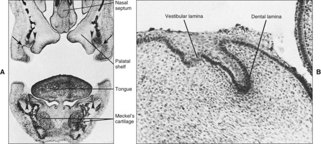
Two or 3 weeks after the rupture of the buccopharyngeal membrane, when the embryo is about 6 weeks old, certain areas of basal cells of the oral ectoderm proliferate more rapidly than do the cells of the adjacent areas. This leads to the formation of the Primary epithelial band which is a band of epithelium that has invaded the underlying ectomesenchyme along each of the horseshoe-shaped future dental arches. At about 7th week the primary epithelial band divides into an inner (lingual) process called Dental lamina and an outer (buccal) process called Vestibular lamina. The dental laminae serve as the primordium for the ectodermal portion of the deciduous teeth. Later, during the development of the jaws, the permanent molars arise directly from a distal extension of the dental lamina.
The development of the first permanent molar is initiated at the fourth month in utero. The second molar is initiated at about the first year after birth, the third molar at the fourth or fifth years. The distal proliferation of the dental lamina is responsible for the location of the germs of the permanent molars in the ramus of the mandible and the tuberosity of the maxilla.
The successors of the deciduous teeth develop from a lingual extension of the free end of the dental lamina opposite to the enamel organ of each deciduous tooth. The lingual extension of the dental lamina is named the successional lamina and develops from the fifth month in utero (permanent central incisor) to the tenth month of age (second premolar).
Fate of dental lamina
It is evident that the total activity of the dental lamina extends over a period of at least 5 years. Any particular portion of the dental lamina functions for a much briefer period since only a relatively short time elapses after initiation of tooth development before the dental lamina begins to degenerate at that particular location.
However, the dental lamina may still be active in the third molar region after it has disappeared elsewhere, except for occasional epithelial remnants. As the teeth continue to develop, they lose their connection with the dental lamina. They later break up by mesenchymal invasion, which is at first incomplete and does not perforate the total thickness of the lamina. Remnants of the dental lamina persist as epithelial pearls or islands within the jaw as well as in the gingiva. These are referred to as cell rest of Serres.
More Confusing Differences in Biology:
Difference Between DNA and RNA
Difference Between Sex and Gender
Difference Between HMO and PPO
Difference Between Dementia and Alzheimer’s
Difference Between Sexual and Asexual Reproduction
Vestibular Lamina
Labial and buccal to the dental lamina in each dental arch, another epithelial thickening develops independently and somewhat later. It is the vestibular lamina, also termed the lip furrow band. It subsequently hollows and forms the oral vestibule between the alveolar portion of the jaws and the lips and cheeks.
As cell proliferation continues, each enamel organ increases in size, sinks deeper into the ectomesenchyme and due to differential growth changes its shape. As it develops, it takes on a shape that resembles a cap, with an outer convex surface facing the oral cavity and an inner concavity.
On the inside of the cap (i.e., inside the depression of the enamel organ), the ectomesenchymal cells increase in number. The tissue appears more dense than the surrounding mesenchyme and represents the beginning of the dental papilla. Surrounding the combined enamel organ and dental papilla, the third part of the tooth bud forms. It is the dental sac or dental follicle, and it consists of ectomesenchymal cells and fibers that surround the dental papilla and the enamel organ.
Thus the tooth germ consists of the ectodermal component-the enamel organ and the ectomesenchymal components-the dental papilla and the dental follicle. The tooth and its supporting structures are formed from the tooth germ. The enamel is formed from the enamel organ, the dentin and pulp from the dental papilla and the supporting tissues namely the cementum, periodontal ligament and the alveolar bone from the dental follicle.
During and after these developments the shape of the enamel organ continues to change. The depression occupied by the dental papilla deepens until the enamel organ assumes a shape resembling a bell. As this development takes place, the dental lamina, which had thus far connected the enamel organ to the oral epithelium, becomes longer and thinner and finally breaks up and the tooth bud loses its connection with the epithelium of the primitive oral cavity.
Development of tooth results from interaction of the epithelium derived from the first arch and ectomesenchymal cells derived from the neural crest cells. Up to 12 days the first arch epithelium retains the ability to form tooth like structures when combined with neural crest cells of other regions. Afterwards this potential is lost but transferred to neural crest cells as revealed in various recombination experiments of first arch ectomesenchyme with various epithelia to produce tooth like structures. Like any other organ development in our body numerous and complex gene expression occurs to control the development process through molecular signals.
In odontogenesis, many of the genes involved or the molecular signals directed by them are common to other developing organs like kidney and lung or structures like the limb. Experimental studies to understand the genetic control and molecular signaling have been done on mice as it is amenable for genetic manipulations like to produce ‘knock-out mice’ ‘or null mice.’ “stages” for descriptive purposes. While the size and shape of individual teeth are different, they pass through similar stages of development. They are named after the shape of the enamel organ (epithelial part of the tooth germ), and are cap, and bell stages.


Leave a Comment
You must be logged in to post a comment.